To solve this problem, researchers at the Karolinska Institute in Sweden are developing DNA repair inhibitors that are able to selectively introduce toxic DNA damage to cancer cells — all while avoiding causing harm to normal cells.
The group’s aim is to support the successful treatment of cancers and improving each patient’s experience of chemotherapy; equipment from Tecan is helping to streamline their research processes and improve their imaging capabilities.
The Helleday Lab is part of the Department of Oncology-Pathology at the Karolinska Institute and primarily investigates basic DNA repair, DNA damage signalling pathways and nucleotide metabolism.
The lab is made up of a multidisciplinary team — including biologists, pharmacologists and medicinal chemists — that work together to support projects all the way from target identification through lead generation and into the clinic. Its aim is to develop novel targeted therapies for cancer, inflammation and autoimmunity, as well as viral infections.
Researchers from the group were among the first to demonstrate the synthetic lethality concept for the treatment of cancer, exploiting the high level of DNA damage and dysfunctional redox status present in cancer cells to prevent the repair of lesions and stimulate further injury to the cell.
The advantage of this approach is that these treatments should have far fewer negative side-effects for patients than existing chemotherapies.
The group is now building on the synthetic lethality concept and expanding its research to target novel DNA repair enzymes.
For example, it has demonstrated how inhibiting MTH1 — a housekeeping enzyme that hydrolyses damaged nucleotides in cells and prevents them from being incorporated into DNA — can specifically target cancer cells without causing toxicity to non-transformed cells.
As a result of this observation, the lab has developed an MTH1 inhibitor — karonudib — which it has progressed through good manufacturing practice (GMP) manufacturing, formulation, good laboratory practice (GLP) safety assessment, and regulatory and ethical approvals, and is now in Phase I clinical trials at Karolinska University Hospital.1–3
Although the main focus within the lab is oncology, the team is also demonstrating the function of its compounds in other applications, such as inflammation, through collaboration with external experts.4
The lab performs a wide range of tests as part of its drug discovery and development workflow, including target engagement assays to ensure that the candidate molecule is binding to the target in the cancer cell, functional assays to test for the efficacy of drugs that induce DNA damage, and synergy assays to investigate how potential anticancer compounds work synergistically with approved drugs.
The group also aims to find out more about the molecular mechanisms of the targets — even if they don’t make it all the way to the clinic — and to develop molecular tools that can be used to find out more about the biology of potential drug targets, which can then be shared with the wider research community.
For all of this work, the team needs to accurately dispense microlitre, nanolitre, or even picolitre volumes of both candidate and approved therapeutics into 384-well plates prior to seeding with cancer cells.
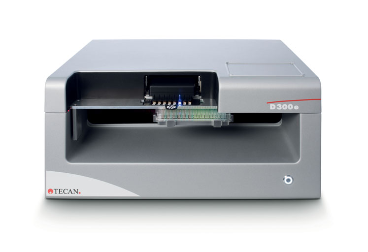
This low volume dispensing is difficult to perform accurately by hand at the scale required; as such, the lab relies on a D300e Digital Dispenser from Tecan to provide non-contact dispensing down to 11 pL.
The D300e is particularly useful for setting up synergy assays in 96- or 384-well formats, which involve the production of complex drug matrixes. Before acquiring the D300e platform, staff had to do these assays manually, making the process extremely time consuming, tedious and prone to human error.
Dr Oliver Mortusewicz, Principal Researcher and Basic Science Team Leader within the group, explained the difference the instrument has made to the lab’s productivity: “The D300e makes our lives so much easier, improving our efficiency and throughput by speeding up our liquid handling workflows and reducing hands-on time. This frees up the staff to do other vital tasks in the lab and reduces the possibility of pipetting errors, which enhances our assay reproducibility.”
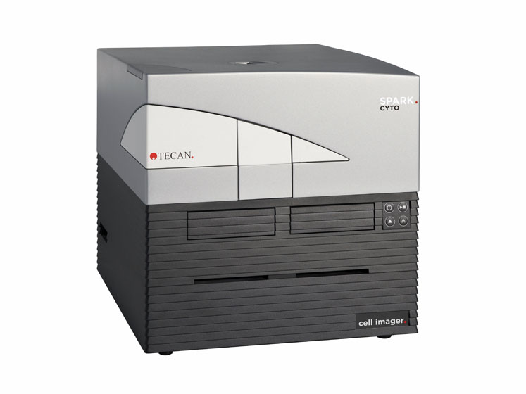
The team is also taking advantage of the real-time imaging and cytometry capabilities of Tecan’s Spark Cyto reader to analyse large numbers of cell viability studies and mechanistic assays.
Oliver continued: “We conduct resazurin assays to test for cell viability, but this test depends on the metabolic activity of the cells, which doesn’t entirely correlate with cell viability. That’s why we subsequently image the plates with our Spark Cyto plate reader.”
“The major advantage of the Spark Cyto compared with a classical plate reader is its ability to perform real-time imaging and cytometry, allowing us to get a better understanding of what is actually happening to the cells."
"For example, we can gauge the rate of proliferation or the localisation of repair proteins upon the induction of DNA damage through the use of fluorescently tagged proteins or immunofluorescence, or perform automated stain-free cell counting and confluence assessments of a whole microplate — requiring just one image per well — using the system’s bright field imaging technology.”
“For mechanistic studies, we can use the Spark Cyto to test our drugs alongside DNA damage-inducing drugs to help us monitor the DNA damage response that we want to target in cancers. By using cells that are fluorescently tagged for a repair protein of interest, we can track the cells following treatment with the damaging agent.”
“We do this by setting up the plates on the D300e and then monitoring responses using the imaging mode of the Spark Cyto. We can also use this method with cell lines that express a specific target or in knockout cell lines that are missing a particular target.”
Oliver and his colleagues now rely on the two Tecan instruments for many of their workflows and are looking forward to employing them in further vital research going forward: “Both of the platforms are so easy to learn and the interfaces are very user friendly,” adds Oliver.
“We have many students working in the lab with us and it doesn’t take long to train them,” he continued: “This means that they can get started on experiments sooner, which is great for our productivity."
"In addition, we have discovered that one of our anticancer molecules is actually also active in preventing inflammation, so we are now venturing out into other areas of research with our drugs, following wherever the science leads us. We certainly plan to keep using our versatile Tecan platforms in the future to support us in our upcoming investigations.”
References
- H. Gad, et al., “MTH1 Inhibition Eradicates Cancer by Preventing Sanitation of the dNTP Pool,” Nature 508(7495), 215–221 (2014).
- K.V. Huber, et al., “Stereospecific Targeting of MTH1 by (S)-Crizotinib as an Anticancer Strategy,” Nature 508(7495), 222–227 (2014).
- U.W. Berglund, et al., “Validation and Development of MTH1 Inhibitors for Treatment of Cancer,” Ann. Oncol. 27(12), 2275–2283 (2016).
- M. Michel, et al., “Small-Molecule Activation of OGG1 Increases Oxidative DNA Damage Repair by Gaining a New Function,” Science 376(6600), 1471–1476 (2022).