Quantitative Western blotting for monitoring biopharma production

Published: 23-Mar-2015

Western blotting methods have changed little in 30 years. But this technique has traditionally been restricted by issues of variability. Mats Falk, GE Healthcare Life Sciences, argues that new technologies and detection techniques herald a new era

You need to be a subscriber to read this article.
Click here to find out more.

Western blotting has been used for more than three decades as an essential tool in the detection, quantification and monitoring of proteins and protein purity. The technique first arose in the late 1970s and was based on the concept for Southern (DNA) blots, developed by Edwin Southern in 1975, and which was followed by Northern (RNA) blotting in 1977. George Stark, Harry Towbin and W. Neal Burnette all contributed to the development of this technique, although it was Burnette who named the technique the ‘Western blot’ after Edwin Southern, in addition to acknowledging Burnette’s US West Coast location.

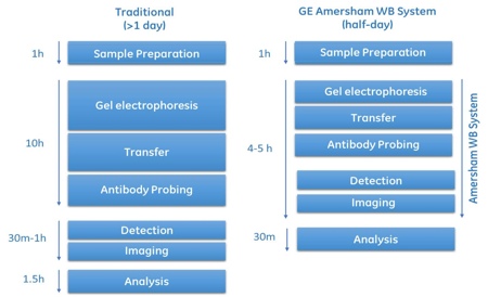
Over the past 30 years, the traditional Western blotting protocol has remained a surprisingly manual technique. A limited number of devices have been available for the automation of individual steps, often with the main focus on speed. Many labs have their own set-up and protocols and some skills are required to obtain high quality results. Western blot workflow involves electrophoresis, transfer, antibody incubation, washing, imaging and analysis of Western blots (Figure 1).

Protein detection techniques

In terms of protein detection, Stark’s group, which also invented the RNA-blotting technique called the ‘Northern blot’, originally used passive transfer of the proteins followed by 125-I-labelled Protein A. This is in contrast to Towbin’s group which, with the knowledge that proteins non-covalently bound to nitro-cellulose, used electrophoretic transfer of proteins to a membrane, then probed with secondary antibodies; this is essentially the procedure still used today.

At present, there are three main detection methods used for Western blotting: chemiluminescence, fluorescence and chemifluorescence

At present, there are three main detection methods used for Western blotting: chemiluminescence, fluorescence and chemifluorescence. With chemi-luminescence, peroxidase is used to catalyse the oxidation of luminol, and in the case of enhanced chemiluminescence, chemical enhancers and catalysts help boost and extend the light emission.

In Western blotting use of horseradish peroxidase (HRP)-conjugated secondary antibodies are the most popular detection technique, where light emitted is proportional to protein concentration. However, developing blots with enhanced chemiluminescence (ECL) has generally remained a manual and time-sensitive procedure, with different X-ray film exposure times being used depending on the signal/protein expression. This has made comparison between different proteins or cell lines difficult, given the confounding variable of different exposure times.

With fluorescence detection, a fluorophore is excited at a particular wavelength, at which point it emits light at another, particular wavelength. The direct source of the fluorescent signal, in addition to the stability of the signal, high sensitivity of detection and the fact that the signal is proportional to the protein concentration, means that fluorescence detection has advantages over chemiluminescence for quantitation. Multiple secondary antibodies conjugated to different fluorophores can also be used for the same blot for fluorescence detection of multiple proteins, when excited at different wavelengths of light.

Another method is chemifluorescence, where alkaline phosphatase (ALP) is reacted with a substrate to produce a fluorescent or chemiluminescent product. Although this method is not as sensitive as chemiluminescence (although more stable), in situations where an ALP-conjugated antibody is available and/or preferred to an HRP-conjugated variety, it is a suitable method for protein detection.

Saturation issues in cases of overloading for detection of low-level proteins can also cause problems

For quantitation it is necessary to ‘normalise’ protein expression either to the total protein content of the samples, or to a ubiquitously-expressed protein (‘housekeeping’ protein). Normalisation versus a housekeeping protein such as GAPDH or actin is commonly used. However, with all three detection methods, the use of a housekeeping protein to normalise the data has proved challenging, as expression of the house-keeping protein can vary based upon the culture conditions, specific cell line, or between different repeats. This can make quantitative data less robust, and comparison of data between different blots more difficult.

Saturation issues in cases of overloading for detection of low-level proteins can also cause problems. In such situations housekeeping product levels might be outside a linear dynamic range for immunodetection, resulting in poor and inaccurate quantification of protein concentration.

Newer normalisation methods using total protein as loading control, such as Cy5 pre-labelling of proteins, gives a total protein concentration and allows normalisation versus total protein, circumventing difficulties with the variability in housekeeping protein expression (Figure 2) and giving more accurate quantitation. It is also easier and more straightforward than the alternative method of post-staining the membrane after transfer to obtain total protein signal for normalisation.

Figure 2: Two of the normalisation methods available with the Amersham WB system

Figure 2: Two of the normalisation methods available with the Amersham WB system

Such consistency in normalisation and fluorescence-based protein detection has transformed Western blotting into a reliable technique with potential applications in the biomanufacturing of therapeutic biologics, where protein expression and purity are paramount.

Apart from Western blot normalisation, Cy5 pre-labelling is sensitive with a broad linear dynamic range and has advantages for SDS-PAGE analysis, compared with traditional protein stains such as silver, Coomassie or SYPRO Ruby. Use of an accurate, sensitive scanner or high quality CCD camera to image the blots after staining (and development, if necessary) is key to accurate analysis.

Quantitative measurement of recombinant protein expression

Use of a fully-integrated Western blotting system that standardises all steps of the traditional blotting process and incorporates Cy5 pre-labelling for total protein concentration, combined with simultaneous detection of target protein with a Cy3 labelled antibody, can facilitate the measurement of reliable quantitative data for biologics production.

In an internal study at GE Healthcare, insect cells were cultured in a single-use rocking bioreactor system for recombinant protein expression. Relative expression levels of (histidine)6-tagged enhanced green fluorescent protein (EGFP) were compared using a monoclonal (histidine)6 antibody, followed by incubation with a polyclonal Cy3B-conjugated secondary antibody. Samples were taken from bioreactor cultures on days 3, 6 and 10. The highest expression was observed 10 days post-infection, with no significant change after this time point (Figure 3). Standardisation of the methodology using the Amersham WB system, along with the minimisation of manual handling and techniques, gave a clear and quantitative measurement of expression levels, and reduced variability in the data.

Figure 3: Expression of (histidine)6-tagged EGFP

Figure 3: Expression of (histidine)6-tagged EGFP

His-tagging is an essential tool used across life sciences research, and involves adding a series of histidine residues to the end of a target protein, for purification with immobilised metal affinity chromatography (IMAC). In another example using histidine-EGFP, target protein recovery was measured on a novel high-productivity metal affinity medium (Capto Chelating). The column was run three times with different concentrations of imidazole in the equilibration and wash buffers, to prevent non-specific binding of endogenous proteins. SDS-PAGE analysis was used to calculate recovery and purity following elution using the Amersham WB system (Figure 4A).

Figure 4a: Expression of (histidine)6-tagged EGFP

Figure 4a: Expression of (histidine)6-tagged EGFP

Monitoring the production and purification of biologics

In monitoring the expression and purity of biologics, use of a robust, quantitative Western blotting system can provide reliable data. In a study at GE Healthcare, quantitative Western blotting was used to measure the expression of an antibody in cell culture media, using both intact (non-reduced) and reduced samples. Looking at protein concentration measurements, both light and heavy chain antibody fragments were detected (Figure 4B). In terms of purity, from 11 days of culture onwards, the intensity of extra bands was observed, most likely from endogenous host cell proteins. Antibody production peaked at around 2mg/ml on day 11. After that, the expression fell and was significantly lower on day 12. A reference product was used for the evaluation and comparison of protein patterns.

Figure 4b: Expression of (histidine)6-tagged EGFP

Figure 4b: Expression of (histidine)6-tagged EGFP

Another example where Western blotting was used to assess the purity of biologics samples in biomanufacturing is the expression of cytokine IFNa-2a. To confirm the identity and purity of the protein, samples were pre-labelled with Amersham WB Cy5 dye labelling reagent and then loaded into the system for Western blotting and analysis. Samples were detected using primary antibodies against IFN a-2a and visualised using Amersham WB goat anti-mouse Cy3 secondary antibodies, as well as scanning for Cy5 signals for total protein by using multiplex imaging capabilities (Figure 5).

Figure 5: Western blot confirming the purity and identify cytokine IFNα-2a, using the Amersham WB system

Figure 5: Western blot confirming the purity and identify cytokine IFNα-2a, using the Amersham WB system

In summary, different protein detection techniques have been used over the years, but for quantitation many favour fluorescence detection’s stability. Fluorescence multiplexing provides the additional advantage of being able to measure multiple proteins in a single blot. Measuring total protein with Cy5 pre-labelling reduces variations seen with endogenous housekeeping proteins. A fully-integrated Western blotting system, with standardised workflow and using fluorescence detection was used to overcome some of the barriers to making this a reliable technique for use in biopharma manufacturing. Recombinant proteins, an antibody and a cytokine were studied, either to find peak production time or analyse concentration, recovery and/or purity following culture and purification steps.

Such applications, combining Cy5 pre-labelling and Cy3 antibody mediated detection in a multiplex analysis, put Western blotting in a strong position to support future biologics manufacturing.

You may also like